

TRIGGER WARNING:obviously this contains gory descriptions. Also related images though these are not super gory!
What is an autopsy?
An autopsy is a procedure carried out to determine cause of death and any contributing factors. It involves a medical examination of a person’s body after they are dead, including externally and internally.
Why would somebody need an autopsy?
In the UK, a death is referred to the coroner if it is determined to be sudden, unexplained, suspicious, violent or unnatural in some way. These include deaths of children, injuries and accidents, people in custody or detained under the Mental Health Act, suicides, drug related deaths, deaths following medical procedures, or other deaths with unknown causes, where a doctor cannot sign off the death certificate with a known cause of death.
An autopsy is also performed at a hospital or specialised mortuary to gain understanding about the death of a patient, to further medical knowledge. This is a hospital autopsy, rather than a forensic autopsy, which is more detailed.
Personnel
The forensic autopsy would be carried out by a forensic pathologist, with the assistance of an anatomical pathology technician. The forensic pathology has many years of medical experience, having been to medical school for 4-5 years, studied the foundation programme for junior doctors for 2 years, and a further histopathology and forensics specialty study of 3 years. There are currently 35-40 Home Office registered forensic pathologists in England and Wales. An anatomical pathology technician undertakes at work training, studying while assisting with autopsies, though these would probably not be the forensic cases until they have more experience or have completed their training courses.
At a forensic autopsy, there may also be others present, including police detectives.
At the Scene
Evaluating the scene is important for the autopsy. This can give context to the findings at autopsy. At a crime scene with a deceased person, the body is the pathologist’s responsibility and the rest of the scene is the forensic investigator’s responsibility. Examining the circumstances surrounding the death can provide important clues to explain any injuries, detail the position of the person as they were injured or killed, the position of the assailant, or other aspects of the recovered body and scene, such as the potential area to search for a bullet that has exited the body.
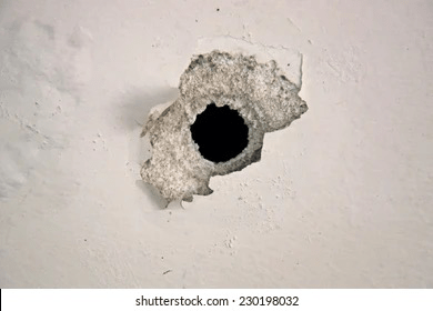
(Image 1)
Autopsy Procedure
Preparation
First, the pathologist must check the paperwork associated with the body they are going to autopsy. The coroner’s form will explain why this death needs an autopsy, and any circumstances surrounding the death. They may look at the medical records of the deceased. Next, the identity of the deceased needs to be matched to the paperwork.
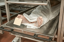
(Image 2)
External Examination
The body to be autopsied would be received into a mortuary, usually at a hospital, in a zip sealed body bag, on a metal gurney. Additional paper bags may have been sealed around the hands if there is potential evidence to be secured, such as under the nails. The body will be stored in a refrigerated container at the mortuary, until such time that it can be autopsied by the forensic pathologist.
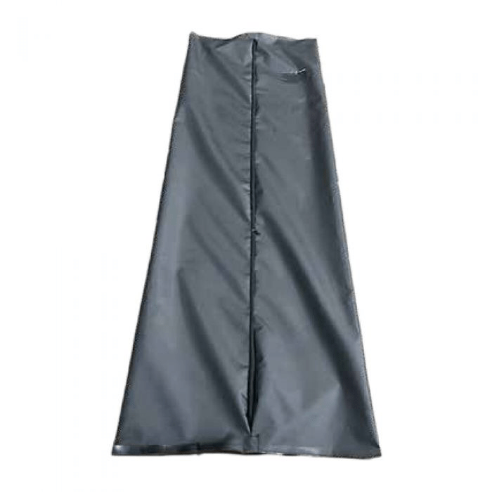
(Image 3)
During the autopsy, the pathologist will verbally record the process, detailing the activities they are undertaking, whilst recording findings on a post-mortem diagram. All photos taken will show a scale and an identification number.
When the autopsy begins, photos may be taken of the body bag including its seal and label. The body bag is opened and the external examination will commence. Full length photos will be taken, usually by standing on a ladder over the body, and include front and back. The body will be weighed and measured for height, also noting sex, ethnicity, age range, and hair and eye colour. Clothing would be removed and the same photos taken again, along with photos of the items removed from the deceased.
Samples of particles or substances on the body will be taken if these are present, and may include paint flakes, plant matter, residues, fibres, scrapings from under the nails and any other foreign material, including a DNA swab if sexual assault is suspected. Samples of hair and nails are taken, and the deceased may be x-rayed or viewed under ultraviolet light to look for any other important details. Then the body will be cleaned, and again full body photos are taken, to document any injuries. Photos of the face will be taken for identification purposes.
The body will be weighed and measured for height, also noting sex, ethnicity, age range, and hair and eye colour. Photos of any injuries will be taken, and any other features of the body such as scars, tattoos, moles, or other marks, as these are found in the external examination. Photos of the hands will be taken also. These may be important for evidence relating to the deceased defending themselves from something or someone who caused their death.
Internal Examination
To begin the internal examination, the deceased is placed on their back with their upper back and shoulders resting on a block, usually made of plastic or rubber. This allows the chest to be pushed upwards for easier incision.
When performing an autopsy, it is important to remember that the deceased may be viewed afterwards by loved ones, and therefore the procedures undertaken during autopsy should be minimally visible when the deceased is dressed again. Taking this into account, the usual incision to access the internal organs of the torso is the Y incision. This begins at each ear, down the side of the neck and around the collarbone, meets at the sternum, and continues down over the abdomen, avoiding the navel and any scars, then ending at the pubic bone.
This incision may be performed by the APT, as well as the subsequent detachment of the abdominal wall tissue from the internal organs with a scalpel. Next, shears are used to cut through the ribs, at the sides of the body, to allow removal of the anterior rib cage – the rib shield – in one piece, once it has been cut from the soft tissue attached to the posterior side of the sternum. This allows for viewing of the heart and lungs in situ before removal.
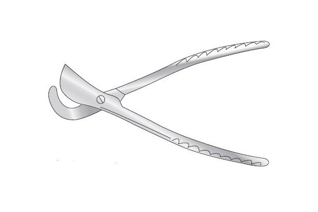
(Image 4)
If there is fluid found in the body cavity, the fluid is removed with special ladles with measurements on, to allow easy measurement of blood or fluid found inside. This will be examined later.
There are different methods used to remove the organs, often a block method – either the entire block of organs, or blocks of multiple organs, cutting the fascia connecting the organs to the interior of the body. The method used varies across the UK. The removal of the organs may sometimes be performed by the APT. The pathologist will then clean and examine the interior of the body, and each organ. Organs are examined, weighed, and cut for a sample or slice to be taken for histological examination. The stomach and intestine contents are examined, weighed, and a sample taken. This would be especially important in cases of poisoning or overdose for example. Major blood vessels are also examined and cut. Photographs can be taken of these processes. The block method may be completed by starting with the neck structures, pulling down the tongue and removing all of the organs together as they are connected. Blunt dissection can be used for this to avoid accidentally cutting any structures.
The Brain
To examine the brain, the head block is moved from under the shoulders to under the head. An incision is made, by the APT, from behind one ear, across the top of the head, ending behind the other ear. Remember that autopsy procedure has to allow for viewing of the deceased if requested after the autopsy. This incision, when sewn back up, is not visible when the deceased is laying on the pillow, especially if they had good hair cover.
The scalp is pulled back from the incision, over the face, and over the back of the neck. The skull will now be visible, and this will be cut with a reciprocating saw, producing a cap-shaped part of the skull which can be removed for access to the brain.
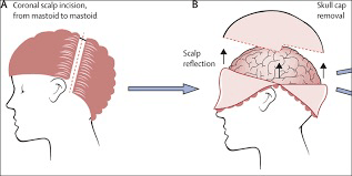
(Image 5)
The brain will be examined in situ, then disconnected from the nerves and spinal cord, to be removed from the skull. To fix the brain for examination or slicing, it will be placed into a container of formalin for 2-4 weeks, otherwise it will have the consistency of jelly, which is not easy to handle or slice.
After the Examination
The body of the deceased must be made suitable for viewing. The internal organs are replaced into the body, inside a bag, to prevent leakage and the cavity is filled with cotton or similar. The tissue and skin that was opened is now sewn back together neatly, remembering the dignity and respect towards the deceased and their family. The skull cap is replaced and the skin is sewn back across the back of the head. The deceased is dressed in a shroud and prepared for viewing. These processes will also be completed by the APT.
Further learning:
Living Autopsy: What Happens During a Post-Mortem? (Full lecture)
https://www.aaptuk.org/link/apt-careers
For information about APTs
REFERENCES:
Image 1 –https://www.shutterstock.com/image-photo/bullet-hole-on-white-wall-230198032
Image 2 – https://en.m.wikipedia.org/wiki/Toe_tag
Image 3 – https://medtree.co.uk/heavy-duty-canvas-body-bag
Image 4 – https://www.surgicalholdings.co.uk/post-mortem/shears/eslander-rib-shear.html
Image 5 –https://gme.uams.edu/wp-content/uploads/sites/24/2018/06/Introduction-to-Autopsy-for-Orientation.pdf
